
Development of an effective and appropriate system for estimating the volume of brain tissue activated for the improvement of therapeutic results in Parkinson’s disease patients that underwent DBS surgery
Deep brain stimulation (DBS) is a surgical technique used primarily to treat movement disorders such as Parkinson’s disease, essential tremor or dystonia, in patients whose symptoms can’t be adequately controlled with medication. The technique involves the application of electrical pulses to a target region in the basal ganglia, in the thalamus or in other subcortical structures by means of an electrode implanted in the brain and connected to a neurostimulator. The adequate adjustment of the neurostimulator parameters is a fundamental step to achieve positive therapeutic results after DBS surgery.
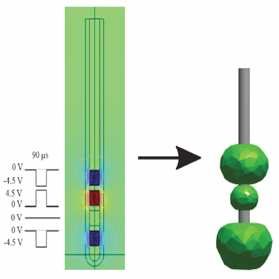
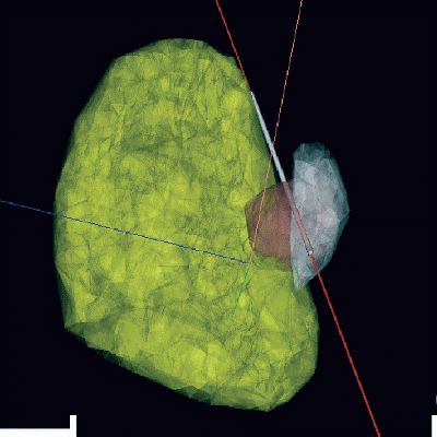
Active tissue volume (VTA) is a metric that represents the extent of direct neuronal activation in response to electrical stimulation. The VTA, as part of a three-dimensional visualization system of the brain, allows the specialist to observe the brain structures that are responding directly to the applied stimulus. Based on this information, the specialist can determine in advance the possible effects, beneficial or harmful, that a given configuration of parameters can have on the patient. Therefore, the adjustment process of the parameters is shortened, saving time and discomfort to the patient. In this project, a VTA estimation system is developed, based on the parameters of stimulation and magnetic resonance imaging (MRI). First, the diffusion tensor of the DBS target regions were calculated from a set of previously filtered DW-WRI images (to cancel noise), and said regions were characterized from the diffusion information obtained. Subsequently, the conductivity tensors were transformed and these results were compared with the conductivity values reported in the literature, obtaining a high similarity.
In second place, a detailed mathematical formulation of the electrostatic process that governs the electrical propagation during the DBS was made, arguing the use of a model based on the Laplace equation. This theoretical analysis was corroborated by a set of computer simulations of the electrical potential generated by the stimulation electrode, carried out using the finite element method. However, it was found that although the quasi-static approach is appropriate from a mathematical perspective, describing the temporal behavior of the input stimulus is fundamental to study the neuronal response to the stimulation (ultimately responsible for the improvement or worsening of the patient’s symptoms). To take into account the temporal characteristics of the electrical stimulation potential, a latent force model was proposed and implemented.
Thirdly, the methodology to calculate the VTA was implemented from the electrical potential generated by the electrode, confirming the variability of the VTA as a function of both the stimulation parameters and the conductivity conditions assumed by the model.
A new approach to reduce the computation time of the VTA based on a Gaussian classifier was also developed, assuming that the VTA calculated from an axon field is equivalent to a binary classification problem.
Fourth, brain structures of interest were segmented from MRI images and DBS electrodes from postoperative CTI, and a specific patient 3D brain atlas was reconstructed.
– VTA estimation system, based on stimulation parameters and magnetic resonance imaging (MRI),
– Approximate calculation of the VTA by Gaussian classifier
Comunications
- Sparse representation of MER signals for localizing the Subthalamic Nucleus in Parkinson's disease surgery. Conf Proc IEEE Eng Med Biol Soc. 2014;2014:950-3.
doi: 10.1109/EMBC.2014.6943749. - A latent force model for describing electric propagation in deep brain stimulation: a simulation study. Conf Proc IEEE Eng Med Biol Soc. 2014;2014:2617-20.
doi: 10.1109/EMBC.2014.6944159. - Electric propagation modeling of deep brain Stimulation (DBS) using the finite element method (FEM). XIX Symposium on Image, Signal Processing and Artificial
Vision (STSIVA), 2014 DOI: 10.1109/STSIVA.2014.7010180 - A Gaussian Process Emulator for Estimating the Volume of Tissue Activated During Deep Brain Stimulation. En IbPRIA 2015: Pattern Recognition and Image Analysis pp
691-699